CPT President Mark Spivak, functioning as a research consultant in conjunction with Emory University’s Neuroscience Department, co-authored a groundbreaking research paper published in the prestigious academic journal PLoS One. The article, entitled Functional MRI in Awake Unrestrained Dogs, coauthored by Dr. Greg Berns, MD, PhD, Andrew Brooks, PhD candidate, and Mark Spivak, CPT, details the first neuroscientific study worldwide that incorporates quality fMRI images in dogs scanned without the use of sedation or restraints.
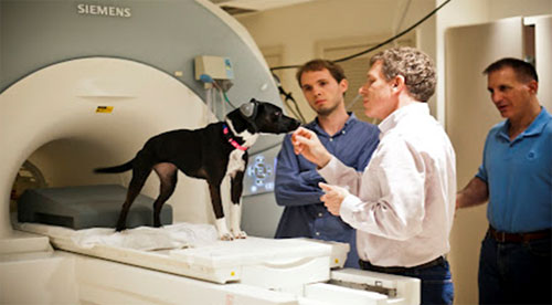
In May 2011, Emory and CPT began collaborating on a humane approach to use functional magnetic resonance imaging (fMRI) to better acquire knowledge related to the human-animal bond, canine cognition, canine emotions, canine sensory perception, and canine receptive communication. Mark co-designed the initial research study, designed the selection criteria for the subject animals, and designed the training protocols, whereby he identified the key variables, then used the processes of systematic desensitization, habituation, behavior shaping, behavior chaining, and positive reinforcement to direct the training of two subject dogs to cooperatively enter the fMRI tube and coil and remain motionless for the required time period.
Until the Emory/CPT research team published the aforementioned PLoS article, the neuroscientific community remained doubtful whether it was possible to effectively scan non-sedated unrestrained animals, as there are a number of pertinent factors that need to be overcome to obtain quality echo planar fMRI images. First, the subject dogs can not move more than 3 mm in any spatial plane for a period of 12 to 25 seconds. Movement in a longitudinal, lateral, vertical, pitch, or yaw direction greater than the referenced distance introduces archetypal noise that destroys the resolution of the image. Furthermore, since fMRI measures brain activity over time, in contrast to a structural MRI that creates internal images of body tissue, once aligned the subject dogs must remain motionless for the duration of each trial repetition. Second, the dogs must willingly enter a narrow cylindrical enclosure (the MRI tube) and then place their head in a more narrow apparatus (the head coil). Consequently, the training process needed to desensitize the dogs to eliminate complications that may arise from potential enclosure anxiety. Third, the dogs must do the preceding while withstanding high-pitched, high-volume noise that varies between 95 to 110 dB. Therefore, the selection process eliminated dogs that exhibited abnormal noise anxiety or noise phobia and the selected dogs were subsequently trained to become comfortable with the fMRI noise using the processes of desensitization and habituation. Fourth, to prevent hearing loss, the dogs had to wear protective ear muffs and wraps. Consequently, the program selection criteria required that the dogs accept handling and grooming, especially about the ear pinna, and the selected dogs were further desensitized to the wearing of the protective equipment. Fifth, the dogs had to perform the required task in an fMRI apparatus designed for the human anatomy. Thus, to minimize anatomical complications, Dr. Berns and Mr. Spivak designed a customized adaptive anatomical foam chin rest that provided greater comfort for the dogs, facilitated static positioning without restraint, and provided greater accuracy when the dogs re-positioning themselves after a reward trial. Sixth, the dogs had to perform their task in an unfamiliar hospital environment. Thus, the selection criteria evaluated for the absence of neophobia, confidence while walking on slippery surfaces, and comportment when working amongst unfamiliar people and dogs. The training protocols further solidified the performance of the selected dogs.
Prior to the Emory/CPT project, when studying animals, neuroscientists used sedation and/or restraints to maintain the static positioning of the subject, usually a primate or rodent. Sedation is acceptable for a structural MRI to scan tissue for veterinary diagnostic purposes. However, for neuroscientific purposes, sedation precludes the researchers from effectively studying cognition. Consequently, when researchers studied awake animals they used mechanical restraints about the head, torso, and extremities, including surgically implanted halos attached to the skull with bolts and cement (the halos were then affixed to the head coil), pillories, stockades, metal braces, and/or leather straps.
In contrast, the Emory/CPT team insisted on using only humane methodologies that studied cognition in an awake pet dog population that participated willingly and cooperatively and that remained in a normal, relaxed emotional state, rather than the anxious emotional state exhibited by the restrained laboratory animals. After obtaining University approval, selecting the first two subject dogs, and training the dogs, the team designed their first study, which measured differences in caudate responses to two human hand-signal communications, one that meant a reward was forthcoming and the other which meant that no reward was forthcoming. The caudate, especially the nucleus accumbens/ventral striatum and to a lesser degree the left amygdala, activate with dopamine and serotonin respectively when there is anticipation of a reward, the expectation of pleasure, or the receipt of pleasure. The study demonstrated that the dogs learned to cognitively and emotionally differentiate the relevance of the two communication signals, as indicated by the significant difference in brain activation dependent upon the particular signal. Moreover, using the humane approach designed by the team, the scan images were very clear. Thus, the team was able to prove to the worldwide neuroscientific community that humane approaches are viable when using fMRI as a tool to study animal cognition.
The groundbreaking methodological advancements achieved by the Emory/CPT team received significant media coverage, including from ABC World New Tonight, ABC News.go.com, the LA Times, Time.com, Rolling Stone.com, Scientific American.com, and Psychology Today. The article about the team was the number one downloaded article on Time.com for a period of 3 weeks. A press release from Emory University was covered by many major and minor media outlets and a YouTube video about the project has received close to 160,000 hits.
The research team has since completed a second study that evaluates the dogs’ emotional response to a variety of canine and human social scents. Planned future research will study canine vision, canine learning, and canine emotions for applications that the group hopes will improve the quality of training protocols for both pet and working purposes. In addition, the Emory/CPT research team plans to expand in July from two subject dogs to 10 trained dogs and then plans to further expand to a team of 30 trained dogs by no later than the first quarter of 2013.
For more information about the Emory/CPT Neuroscience Project, please review the links contained in this article or contact Mark Spivak at CPT by email (MarkCPT@aol.com) or by phone (404-236-2150).