
CPT and Dog Star Technologies founder Mark Spivak, along with Gregory Berns, MD, PhD, Department of Psychology, Emory University; Sarah Nemanic, DVM, PhD, Carlson College of Veterinary Medicine, Oregon State University; and Nicole Northrup, DVM, DACVIM (Oncology), College of Veterinary Medicine, University of Georgia, collaborated to author the paper “Clinical Findings in Dogs Trained for Awake-MRI” published in the most recent edition of Frontiers in Veterinary Science. The paper discusses case studies and practical veterinary applications produced via the novel awake canine-MRI process developed by Berns and Spivak.
Training dogs for awake-MRI and fMRI began in 2012 for the study of canine cognition, emotions, sensory perception, and receptive communication. Although originally envisioned principally as a research technique to understand the neuro-behavioral mechanisms of the canine brain, its significant potential as a new diagnostic tool has become apparent.

Initially, the versatility of the methodology was recognized serendipitously, when a subject dog participating in an Emory/CPT study funded by the Office of Naval Research was found to have hydrocephalus during an adjunct structural MRI of the dog’s brain. Berns and Spivak then realized that they may have stumbled upon an innovative veterinary resource for diagnosing central nervous system disease. They later modified the structural scanner sequences to reduce noise, while improving image quality and acquisition speed. They continued advancing the technique to acquire high resolution images of visceral organs, which provides the opportunity to detect abdominal cancer, such as fast metastasizing splenetic, hepatic, and pancreatic hemangiosarcomas, during early stages, when veterinary intervention has a higher probability of significantly impacting patient longevity.
The methodology created by Berns and Spivak, which is available to pet owners and veterinarians through Dog Star Technologies, completes a head/neck structural scan in 26 – 30 seconds and an abdominal scan in only 90 seconds. Moreover, the images are acquired without the use of anesthesia on a high-powered 3-Tesla MRI scanner.
In comparison, the preceding scans on a .5T scanner at a typical veterinary specialty hospital require anesthesia and occupy 1.5 – 2.5 hours of sedated patient time. Furthermore, because of the extensive scanner, labor, and anesthesia hours the cost is generally between $2,000 – $2,200, whereas Dog Star Technologies is projecting a cost of only $400 – $600 for a comparable awake-MRI scan.
The paper presented 4 case examples of canine patients, where without anesthesia and without exposure to ionizing radiation in less than 5 minutes awake MRI effectively diagnosed: 1) nasal carcinoma, 2) brain tumor, 3) abdominal lipoma, and 4) idiopathic epilepsy.
Participating dogs received 2 –4 months of awake MRI training. A human knee coil was used for head scans. The bore’s body coil was used for visceral scans. Using a 3T Siemens Trio MRI scanner and either T2-weighted or BLADE imaging sequences Berns/Spivak obtained high-resolution structural scans pertinent to each diagnosis.
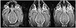
In the first case, an 8-year old Bouvier des Flandres was experiencing a nosebleed of unknown origin. A physical exam by a local veterinarian detected no abnormalities. However, in less than 30 seconds awake MRI detected a large neoplastic mass in the upper sinus. The observation of Berns/Spivak was confirmed by one of the paper’s co-authors, Nicole Northrup, a board-diplomated veterinary oncologist at the University of Georgia Veterinary School. Dr. Northrup subsequently treated the dog with targeted radiation. During the treatment period Berns/Spivak used awake MRI to monitor the tumor suppression achieved from the dog’s treatment.
In the second case, a 12-year old Golden Retriever was experiencing seizure activity. Awake MRI detected a frontal lobe mass that was the likely cause of the epileptic events. The observation of Berns/Spivak was corroborated by one of the paper’s co-authors, Sarah Nemanic, a veterinary radiologist at the Carlson College of Veterinary Medicine, Oregon State University.

In the third case, a 10-year Pit Bull/Vizsla exhibited external cystic masses. Awake MRI identified corresponding subcutaneous abdominal tumors, later identified as lipomas. The image was acquired by Berns/Spivak and analyzed by Dr. Nemanic.
In the fourth case, a 3-year old Boxer/Hound was experiencing experiencing epileptic seizures of increasing frequency and severity. Awake MRI detected ventriculomegaly that was a potential cause of the seizure activity. The Berns/Spivak observation was confirmed by Dr. Nemanic. The owner then took the MRI film to her dog’s veterinary neurologist to discuss whether such knowledge should prompt an adjustment in the treatment regimen.
In the opinion of the authors, awake MRI provides potentially significant benefit as a health monitoring tool for breeds at high risk for brain and/or abdominal cancer, including popular breeds such as Golden Retrievers, Labrador Retrievers, German Shepherds, Boxers, and Collies. Furthermore, awake MRI has utility as a diagnostic tool in cases of canine epilepsy. If dogs are trained at a young age to obtain baselines, then subsequently scanned 1 –4 times/yr., depending upon age, the authors believe that the technique can likely detect cancer at much earlier ages, which will allow more successful veterinary intervention.

Persons wishing to learn more about the process, marketed through Dog Star Technologies as “Health Detector,” should contact Mark Spivak via email (MarkCPT@aol.com) or phone (404-236-2150).
Atlanta, GA
Sandy Springs, Ga
Decatur, GA

Need to get in touch with CPT? I’ve got you covered.
Pick what works best for you!


